
Why PET/CT Specifications Matter More Than You Think
A PET/CT scanner represents one of the most significant capital investments a healthcare facility can make. Whether you are evaluating a new system or a pre-owned alternative, the specifications listed on a datasheet directly influence your clinical outcomes, diagnostic confidence, and long-term return on investment. However, not all specifications are created equal — and not all of them are measured the same way across manufacturers.
Therefore, understanding the standards behind the numbers is just as important as the numbers themselves. Three specification categories consistently surface during PET/CT evaluations: NEMA performance standards, system sensitivity, and spatial resolution. Each tells you something different about how a scanner will perform in your clinical environment, and together, they form the foundation of an informed purchasing decision.
What Are NEMA Standards in PET/CT Imaging?
The National Electrical Manufacturers Association (NEMA) publishes a standardized testing protocol — known as NEMA NU 2 — that defines exactly how PET scanner performance should be measured and reported. This protocol exists for one critical reason: to give you an apples-to-apples comparison between different PET/CT systems, regardless of the manufacturer.
Without NEMA NU 2, every Original Equipment Manufacturer (OEM) could measure sensitivity, resolution, and count rate performance using its own methodology. The result would be marketing-driven numbers that are nearly impossible to compare across brands. NEMA NU 2 eliminates that problem by prescribing specific phantoms, acquisition protocols, and data analysis methods that every manufacturer must follow when publishing performance claims.
Key NEMA NU 2 Performance Metrics
The NEMA NU 2 standard covers several performance characteristics, but the ones most relevant to your purchasing decision include the following:
- Spatial resolution — measures the scanner's ability to distinguish between two closely spaced structures.
- Sensitivity — measures the system's ability to detect coincidence events from a given radiotracer activity.
- Scatter fraction and count rate performance — evaluates how well the scanner handles high-activity imaging scenarios without signal degradation.
- Image quality — assessed using a standardized phantom that simulates clinical lesion detection tasks.
- Timing resolution — particularly important for time-of-flight (TOF) PET systems, where faster timing translates to improved signal-to-noise ratios.
Simply put, NEMA NU 2 gives you a reliable, standardized framework for evaluating any PET/CT system — whether it is a current-generation model from Siemens Healthineers, GE Healthcare, Philips, or Canon Medical.
PET/CT Sensitivity: Detecting What Matters
Sensitivity is one of the most consequential specifications on any PET/CT datasheet. It tells you how efficiently the scanner converts radiotracer decay events into usable imaging data. A system with higher sensitivity detects more coincidence photon pairs per unit of injected activity, which has direct implications for both image quality and patient dose.
NEMA NU 2 measures sensitivity in counts per second per kilobecquerel (cps/kBq). The test uses a standard line source placed at the center and at a 10 cm offset from the center of the scanner's axial field of view (FOV). The results quantify how much of the emitted radiation the detector ring actually captures.
Why Higher Sensitivity Matters Clinically
Higher sensitivity creates a cascade of clinical advantages. When the system detects more events, you can achieve diagnostic-quality images with shorter scan times, lower injected doses, or both. For oncology departments managing high patient throughput, this translates to more scans per day without compromising diagnostic confidence.
Furthermore, higher sensitivity is particularly valuable in pediatric imaging and in facilities that perform serial follow-up studies. Reducing the injected dose per exam lowers cumulative radiation exposure — a growing concern among referring physicians and patients alike.
What Drives Sensitivity Differences Between Systems?
Several hardware design factors determine a PET/CT system's sensitivity. The most influential include:
- Detector material — Lutetium-based scintillators (such as LSO and LYSO) offer superior stopping power and light output compared to older BGO crystals, directly increasing detection efficiency.
- Axial field of view — A longer axial FOV means more detector rings, which capture a greater solid angle of emitted photons. Systems with extended axial coverage — such as those exceeding 25 cm — demonstrate markedly higher sensitivity than standard 15–18 cm configurations.
- Detector ring geometry — The diameter and arrangement of the detector ring influence geometric efficiency. Tighter ring diameters increase the solid angle coverage but must be balanced against patient bore size requirements.
On the other hand, sensitivity numbers alone do not tell the full story. A system with excellent raw sensitivity but poor timing resolution or high scatter fraction may not deliver the image quality improvement you expect. This is why evaluating specifications in combination — rather than in isolation — is essential.
Spatial Resolution: The Detail Behind the Image
Spatial resolution defines the smallest structure a PET/CT scanner can reliably distinguish. In practical terms, it determines whether a 4 mm lesion appears as a distinct focus of uptake or blurs into the surrounding tissue. For oncology staging, radiation therapy planning, and treatment response assessment, spatial resolution directly impacts clinical decision-making.
NEMA NU 2 measures spatial resolution using point sources placed at defined positions within the scanner's FOV. The results are reported as full width at half maximum (FWHM) values in millimeters, measured in both the transverse and axial planes, at the center and at a 10 cm radial offset. Lower FWHM values indicate finer spatial resolution.
Factors That Influence Spatial Resolution
Spatial resolution in PET imaging is governed by a combination of physical and engineering factors. Understanding these helps you interpret datasheet values with greater precision.
- Crystal size — Smaller individual detector crystals improve intrinsic spatial resolution. Modern systems use crystals as small as 3–4 mm, compared to 6 mm or larger in older-generation scanners.
- Positron range — This is a physics-based limitation that depends on the radiotracer. Fluorine-18 (F-18), the most commonly used PET isotope, has a relatively short positron range, which supports better spatial resolution compared to tracers like Rubidium-82.
- Photon non-collinearity — The two annihilation photons are not emitted at exactly 180 degrees, introducing a small but unavoidable angular uncertainty. This effect worsens with larger detector ring diameters.
- Reconstruction algorithms — Iterative reconstruction methods, particularly those incorporating point spread function (PSF) modeling, can recover spatial resolution lost during acquisition.
Spatial Resolution at Center Versus Offset
An important nuance to understand is that spatial resolution typically degrades as you move away from the center of the FOV. NEMA reports values at both center and 10 cm offset for exactly this reason. A scanner may advertise an impressive 4 mm FWHM at center, but perform at 5.5 mm or worse at the periphery.
This matters clinically because many structures of interest — axillary lymph nodes, peripheral lung lesions, pelvic masses — are not located at the geometric center of the scanner. Therefore, evaluating off-center resolution gives you a more realistic picture of everyday diagnostic performance.
How Sensitivity and Spatial Resolution Work Together
It can be tempting to focus on a single metric when comparing PET/CT systems. However, sensitivity and spatial resolution interact in ways that directly affect your clinical experience. A system with outstanding sensitivity but mediocre spatial resolution will produce statistically robust images that lack fine anatomical detail. Conversely, a system with excellent spatial resolution but low sensitivity may require longer scan times or higher injected doses to achieve acceptable image quality.
The most clinically capable systems balance both specifications effectively. Modern PET/CT scanners from leading OEMs achieve this balance through a combination of advanced scintillator materials, optimized crystal geometry, and sophisticated reconstruction algorithms that incorporate both TOF and PSF correction data.
Nevertheless, the ideal balance depends on your specific clinical requirements. A high-volume oncology center performing primarily whole-body FDG studies may prioritize sensitivity and throughput. A research-oriented facility focused on neuroimaging or small-lesion characterization may place greater weight on spatial resolution. Neither approach is wrong — what matters is that your specifications align with your clinical mission.
Evaluating PET/CT Specifications on Pre-Owned Systems
When you consider a pre-owned PET/CT scanner, the NEMA-published specifications remain your most reliable reference point. These values reflect the system's design performance, not its age. A well-maintained pre-owned system will perform at or near its original NEMA benchmarks, provided the detector block integrity and calibration are properly maintained.
However, several practical factors deserve your attention during the evaluation process:
- Detector crystal condition — Over time, individual detector elements can degrade or fail. A quality pre-owned system should include documentation of any dead or underperforming detector blocks.
- Calibration history — Regular normalization and calibration ensure that the system's measured performance matches its design specifications. Request calibration records when evaluating any pre-owned PET/CT.
- Software version — Reconstruction capabilities, including TOF and PSF modeling, depend on the software revision. Verify that the system runs a software version that supports the advanced reconstruction features advertised in its original NEMA data.
- CT component condition — A PET/CT is a dual-modality system. The CT subsystem's tube hours, detector condition, and software version are equally important to evaluate.
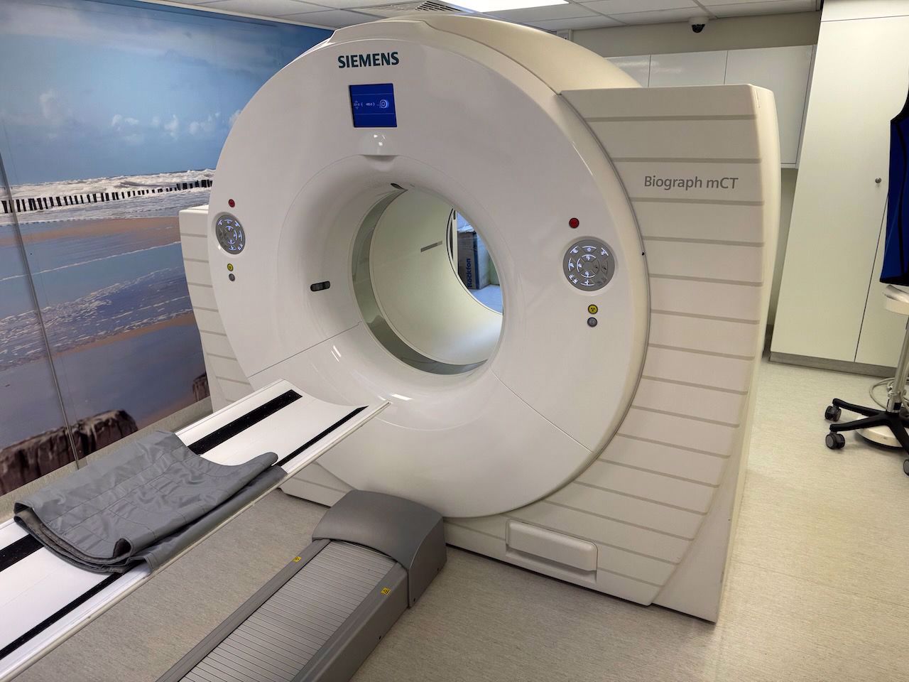
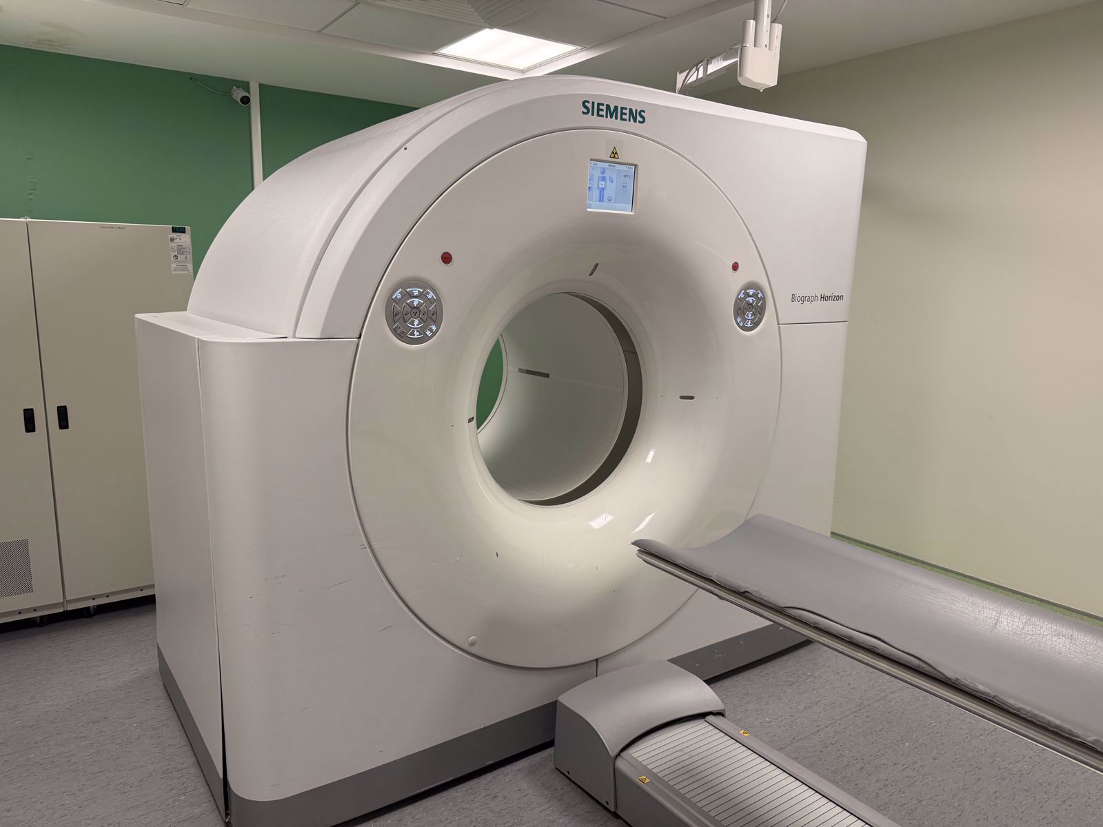
At Scandinavian Medical Solutions, we source pre-owned PET/CT systems from all four major OEM brands — Siemens Healthineers, GE Healthcare, Philips, and Canon Medical. Our team evaluates every system against its original technical specifications, so you receive equipment that meets OEM standards and aligns with your clinical requirements.
Beyond the Datasheet: Matching Specifications to Your Clinical Workflow
Specifications provide the technical foundation, but your purchasing decision should ultimately be driven by how those specifications translate into your daily clinical workflow. A system with a 25 cm axial FOV and high sensitivity may be ideal for a busy community hospital performing 15 to 20 PET/CT studies per day. A system with finer spatial resolution and advanced cardiac gating capabilities may better serve a specialized cardiac imaging center.
Furthermore, operational considerations — such as room dimensions, power supply requirements, helium consumption (for MRI-compatible hybrid installations), and ongoing service support — should factor into your decision alongside NEMA performance data. A technically superior system that does not fit your physical space or service infrastructure creates more problems than it solves.
This is where working with an independent partner provides a distinct advantage. Because we are not tied to a single manufacturer, we help you evaluate PET/CT options across brands and generations based entirely on your clinical requirements and operational constraints — not on a sales quota for a specific product line.
Summary
PET/CT specifications are not just numbers on a page — they are the measurable indicators of a system's clinical capability. NEMA NU 2 standards provide you with a reliable, manufacturer-independent framework for comparing system sensitivity, spatial resolution, count rate performance, and image quality. Sensitivity determines how efficiently a scanner collects data, directly affecting scan time, patient dose, and throughput. Spatial resolution determines the level of anatomical detail visible in your images, influencing lesion detection and staging accuracy.
The most effective approach is to evaluate these specifications together, in the context of your facility's specific clinical mission and operational realities. Whether you are acquiring a new system or a pre-owned alternative, understanding what the numbers mean — and how they were measured — puts you in a stronger position to make the right decision.
We Are Here to Help You Find the Right PET/CT System
Whether you are evaluating PET/CT specifications for an upcoming purchase, need spare parts to maintain an existing system, or require a flexible rental solution to bridge a gap in your imaging capacity, we are here for you. At Scandinavian Medical Solutions, we offer pre-owned PET/CT systems and spare parts from GE Healthcare, Siemens Healthineers, Philips, and Canon Medical — all evaluated against OEM standards.
Do not hesitate to contact our team for guidance in finding the right system for your clinical requirements. You can reach us at +45 5080 8009 or +1 (714) 240-0864 or email us at sales@scandinavian-medical.com. We are your trusted partner every step of the way.