
Why the CT Component Matters More Than You Think in PET/CT
The CT subsystem inside a PET/CT scanner is far more than an anatomical backdrop for metabolic imaging. It directly shapes diagnostic accuracy, patient dose, and the overall clinical utility of every scan your facility performs. When the CT component underperforms — whether due to aging hardware, suboptimal protocols, or outdated reconstruction software — the consequences ripple through attenuation correction, lesion localization, and radiation exposure.
Therefore, understanding how CT integration affects both image quality and dose management is essential for any radiology department evaluating a new or pre-owned PET/CT system. The decisions you make about the CT side of the equation will influence clinical outcomes, regulatory compliance, and long-term operational costs for years to come.
The Dual Role of CT in PET/CT Imaging
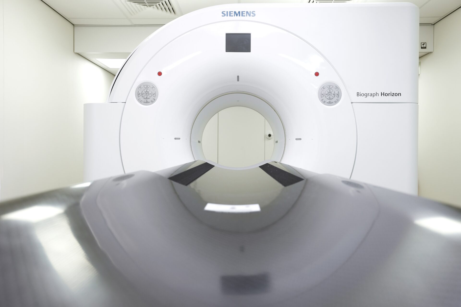
A PET/CT scanner combines two distinct imaging modalities into a single gantry. The PET (Positron Emission Tomography) component detects gamma rays emitted by a radiotracer — typically fluorodeoxyglucose (FDG) — to reveal metabolic activity. The CT (Computed Tomography) component provides high-resolution anatomical images and generates the attenuation correction map that PET data requires for quantitative accuracy.
Simply put, without a properly functioning CT subsystem, even the most advanced PET detector ring cannot produce reliable diagnostic images. The CT data corrects for the way gamma photons are absorbed and scattered by different tissue densities in the patient's body. If the CT attenuation map is noisy, misaligned, or acquired at inappropriately low settings, the resulting PET images will contain quantitative errors that can affect clinical decision-making.
Attenuation Correction: The Foundation of Quantitative PET
Attenuation correction (AC) converts raw PET emission data into standardized uptake values (SUVs) that oncologists, cardiologists, and neurologists rely on for diagnosis and treatment monitoring. The CT scan acquired during the PET/CT examination generates a pixel-by-pixel map of tissue density, which the reconstruction algorithm uses to compensate for photon attenuation.
When the CT component delivers consistent, artifact-free images, the AC map is accurate — and SUV measurements become reproducible across serial studies. However, when the CT data contains beam-hardening artifacts from metal implants, truncation artifacts from a limited field of view, or excessive noise from low-dose protocols pushed too far, the AC map degrades. This can lead to false-positive or false-negative findings in oncologic staging, therapy response assessment, and surveillance imaging.
Anatomical Localization and Diagnostic CT
In many clinical scenarios, the CT component of a PET/CT study serves a dual diagnostic purpose. Rather than acquiring a low-dose CT solely for attenuation correction, facilities often run a full diagnostic-quality CT with intravenous contrast during the same session. This approach eliminates the need for a separate CT appointment, reduces overall radiation burden, and streamlines the patient workflow.
For this diagnostic CT capability to deliver genuine clinical value, the CT subsystem must offer sufficient slice count, spatial resolution, and dose modulation technology. A 16-slice CT integrated into an older PET/CT platform, for example, may struggle to produce the temporal resolution needed for cardiac PET/CT or the thin-slice reconstructions required for pulmonary nodule characterization. On the other hand, a 64-slice or 128-slice CT subsystem opens the door to a much broader range of diagnostic applications within the PET/CT workflow.
Dose Management in PET/CT: Balancing Quality and Safety
Radiation dose in PET/CT comes from two sources: the injected radiotracer and the CT acquisition. While the PET dose is largely fixed by the administered activity, the CT dose is highly variable and directly controllable through protocol optimization. In many studies, the CT component accounts for 50% or more of the total effective dose delivered to the patient.
This makes CT dose management one of the most impactful areas where your team can improve patient safety without sacrificing diagnostic performance. Furthermore, regulatory scrutiny around cumulative radiation exposure continues to increase — particularly for pediatric patients and adults undergoing serial PET/CT examinations for cancer surveillance.
Automatic Tube Current Modulation
Automatic tube current modulation (ATCM) is one of the most effective dose-reduction tools available on modern CT subsystems. ATCM adjusts the milliampere (mA) output in real time based on the patient's anatomy — increasing current through denser structures like the pelvis and reducing it through less attenuating regions like the lungs.
The sophistication of the ATCM algorithm varies significantly between CT generations and manufacturers. Newer systems from GE Healthcare, Siemens Healthineers, Philips, and Canon Medical (formerly Toshiba) implement both angular and longitudinal modulation, which can reduce dose by 20–40% compared to fixed-mA techniques while maintaining diagnostic image quality. When evaluating a PET/CT system — whether new or pre-owned — confirming the ATCM capabilities of the CT subsystem is an essential step.
Iterative Reconstruction and Its Impact on Dose
Iterative reconstruction (IR) algorithms have transformed CT dose management over the past decade. Unlike traditional filtered back projection (FBP), which amplifies noise at lower dose levels, IR algorithms use statistical modeling to reduce image noise while preserving spatial resolution and contrast. This allows you to lower the tube current — and therefore the dose — without the corresponding increase in image noise that would occur with FBP alone.
Each major Original Equipment Manufacturer (OEM) offers proprietary IR solutions: Siemens provides ADMIRE, GE offers ASiR-V, Philips uses iDose⁴ and IMR, and Canon Medical developed AIDR 3D. The generation and type of IR available on a given PET/CT system depends on the CT subsystem's hardware and software platform. Therefore, when assessing a pre-owned PET/CT scanner, verifying the installed reconstruction software — and whether it can be upgraded — is a critical technical consideration.
Low-Dose CT Protocols for Attenuation Correction
When the CT component is used solely for attenuation correction and anatomical localization — without a diagnostic intent — significantly lower dose settings can be employed. Many facilities run their AC-only CT at 80–120 kVp with reduced mA settings, resulting in effective doses as low as 1–3 mSv.
Nevertheless, it is important to remember that reducing dose too aggressively can introduce excessive noise into the attenuation map, which in turn propagates errors into the corrected PET images. Finding the right balance between dose reduction and AC map quality requires careful protocol optimization — and a CT subsystem capable of delivering stable performance at low-dose settings. Older X-ray tubes with high accumulated scan seconds or approaching end of life may struggle to maintain consistent output at these lower operating parameters.
Key CT Specifications That Drive PET/CT Performance
Not all CT subsystems integrated into PET/CT scanners are created equal. The CT specifications directly determine the range of clinical applications your PET/CT can support, the dose efficiency you can achieve, and the longevity of the system before major component replacements become necessary. Here are the specifications that matter most.
Slice Count and Detector Configuration
The number of detector rows — commonly referenced as the slice count — defines the CT subsystem's volumetric coverage per rotation. A 16-slice CT covers a narrower z-axis per rotation than a 64-slice or 128-slice system, which means longer scan times, more rotations, and potentially higher cumulative dose for whole-body PET/CT acquisitions.
Higher slice counts also enable thinner reconstruction slices, which improve spatial resolution for small-lesion detection and allow multiplanar reformats that match the quality of a standalone diagnostic CT. For facilities that intend to use the CT component for diagnostic purposes — not just attenuation correction — a 64-slice CT subsystem or higher is generally recommended.
X-Ray Tube Capacity and Life Expectancy
The CT X-ray tube is the most critical — and most expensive — consumable component in any CT-based imaging system. Its capacity, typically measured in Mega Heat Units (MHU), determines how much thermal stress it can absorb before requiring a cooling period or replacement. High-throughput PET/CT facilities that perform 15–25 studies per day place considerable thermal demands on the tube.
When evaluating a pre-owned PET/CT scanner, understanding the tube's remaining life expectancy is essential. Key usage metrics — including total scan seconds, total milliampere-seconds (mAs), and total patient examinations — provide a comprehensive picture of how much operational life remains. We always recommend requesting these tube usage statistics before any purchase decision, as tube replacement can represent a significant portion of the system's total cost of ownership.
Gantry Bore Size and Patient Comfort
PET/CT gantry bore diameter typically ranges from 70 cm to 78 cm, depending on the model and generation. A wider bore accommodates a broader range of patient body habitus, improves comfort during the relatively long PET/CT acquisition, and reduces the likelihood of scan truncation artifacts in the CT field of view — which, as discussed earlier, can compromise attenuation correction accuracy.
For radiation therapy planning applications, where PET/CT is increasingly used for target volume delineation, a wide-bore configuration with a flat table top is particularly important. These considerations should inform your system selection process regardless of whether you are acquiring a new or pre-owned unit.
Evaluating CT Quality in Pre-Owned PET/CT Systems
Purchasing a pre-owned PET/CT scanner offers a cost-effective path to advanced hybrid imaging — but only when you evaluate the CT subsystem with the same rigor you would apply to a standalone CT purchase. The PET detector ring tends to age more gracefully than the CT side, where mechanical components like the X-ray tube, slip rings, and cooling system experience continuous wear.
A thorough evaluation should include a review of the CT tube usage data, a verification of the installed software version and available dose-reduction features, confirmation of the detector calibration status, and an assessment of the gantry mechanical condition. Furthermore, understanding what spare parts are available for the specific CT subsystem model — and their lead times — helps you project ongoing maintenance costs with accuracy.
At Scandinavian Medical Solutions, we specialize in helping facilities navigate these technical complexities. Our team evaluates every PET/CT system in our inventory against OEM standards, and we provide transparent documentation of CT tube metrics, software configurations, and system histories. We believe that an informed buyer makes the best long-term decisions — and we are here to ensure you have every piece of information you need.
Maintaining Diagnostic Quality and Dose Efficiency Over Time
CT integration in PET/CT is not a one-time consideration. Maintaining diagnostic quality and dose efficiency requires ongoing attention to calibration, protocol optimization, and timely component replacement. Regular quality control — including daily air calibrations, periodic phantom scans, and annual physicist reviews — ensures that the CT subsystem continues to deliver consistent Hounsfield unit accuracy and acceptable noise levels.
Proactive tube monitoring is equally important. Tracking the tube's accumulated mAs and scan seconds against the manufacturer's recommended thresholds allows your biomedical engineering team to plan replacements before unexpected failures disrupt your clinical schedule. Access to reliable spare parts — from X-ray tubes and high-voltage generators to detector modules and cooling system components — determines how quickly you can restore full functionality when maintenance events arise.
We maintain an inventory of over 5,000 unique medical imaging spare parts from all major OEM brands, including Siemens Healthineers, GE Healthcare, Philips, and Canon Medical. Every part we supply undergoes rigorous testing and is shipped with secure, purpose-designed packaging to ensure it arrives ready for installation.
Your Trusted Partner for PET/CT Equipment and Support
Whether you are evaluating a pre-owned PET/CT system for your facility, seeking a replacement CT X-ray tube, or looking for guidance on optimizing your current scanner's dose protocols, we are here for you. At Scandinavian Medical Solutions, we combine deep technical knowledge with a consultative approach — because we understand that every imaging decision you make carries real clinical and financial weight.
Our team offers pre-owned PET/CT scanners from GE, Siemens, Philips, and Canon Medical, along with comprehensive spare parts support and flexible rental solutions for facilities that need interim capacity. We do not simply sell equipment — we help you find the right solution for your specific clinical requirements.
Do not hesitate to reach out to our team for a consultation. You can call us at +45 5080 8009 or +1 (714) 240-0864 or email us at sales@scandinavian-medical.com. We are ready to help you secure the diagnostic quality and dose efficiency your patients and your department deserve.