
Why PET/CT Technology Matters in Modern Diagnostics
Few imaging modalities have transformed oncology, cardiology, and neurology as profoundly as the PET/CT scanner. By combining two powerful diagnostic technologies into a single system, PET/CT gives clinicians the ability to see both the structure and the metabolic activity of tissues—simultaneously and with remarkable precision.
For hospitals and imaging centers evaluating their next equipment investment, understanding how a PET/CT scanner works—and what distinguishes one system from another—is not optional. It is essential. The financial commitment is significant, and the clinical impact on patient outcomes is even greater.
Therefore, this article breaks down the core principles, key components, and distinct advantages of PET/CT scanners. Whether you are a radiology department head planning a new installation or a biomedical engineer evaluating pre-owned systems, the information here will help you make more confident decisions.
The Fundamental Principles Behind PET/CT Imaging
A PET/CT scanner integrates two distinct imaging modalities into one gantry. Each modality contributes different—but complementary—diagnostic information. To fully appreciate the value of the combined system, you need to understand each one individually.
Positron Emission Tomography (PET): Functional Imaging
PET imaging works by detecting gamma rays emitted from a radiotracer injected into the patient's body. The most commonly used radiotracer is fluorodeoxyglucose (FDG), a glucose analog that accumulates preferentially in cells with high metabolic activity—such as cancer cells.
Once the radiotracer decays, it emits positrons. Each positron quickly collides with an electron, producing two gamma photons that travel in opposite directions. The PET detector ring surrounding the patient captures these photon pairs, and the system's reconstruction algorithms calculate the precise origin of each emission event.
Simply put, PET reveals how tissues are functioning at a cellular level. It answers the question: "Is this tissue metabolically active, and to what degree?" This makes PET exceptionally powerful for detecting tumors, evaluating treatment response, and identifying recurrence.
Computed Tomography (CT): Anatomical Imaging
CT imaging uses an X-ray tube rotating around the patient to generate cross-sectional images of the body. The detector array on the opposite side of the gantry captures the attenuated X-rays, and the system reconstructs detailed anatomical images based on density differences in tissue.
CT excels at providing high-resolution structural detail. It shows you exactly where an abnormality is located, its size, and its relationship to surrounding anatomy. However, CT alone cannot reliably distinguish between a metabolically active lesion and a benign structural change.
This is precisely where the combination of PET and CT becomes so valuable.
The Power of Fusion: PET and CT Together
When PET and CT data are acquired in the same scan session and fused by the system's software, the result is an image that overlays metabolic activity onto a precise anatomical map. The clinician can see not only that abnormal metabolism exists, but exactly where it is occurring within the patient's body.
Furthermore, the CT data serves a dual purpose in PET/CT systems. Beyond providing anatomical reference, it is used for attenuation correction—a mathematical process that compensates for the way gamma photons are absorbed or scattered as they pass through different tissue types. This correction significantly improves the quantitative accuracy of the PET images.
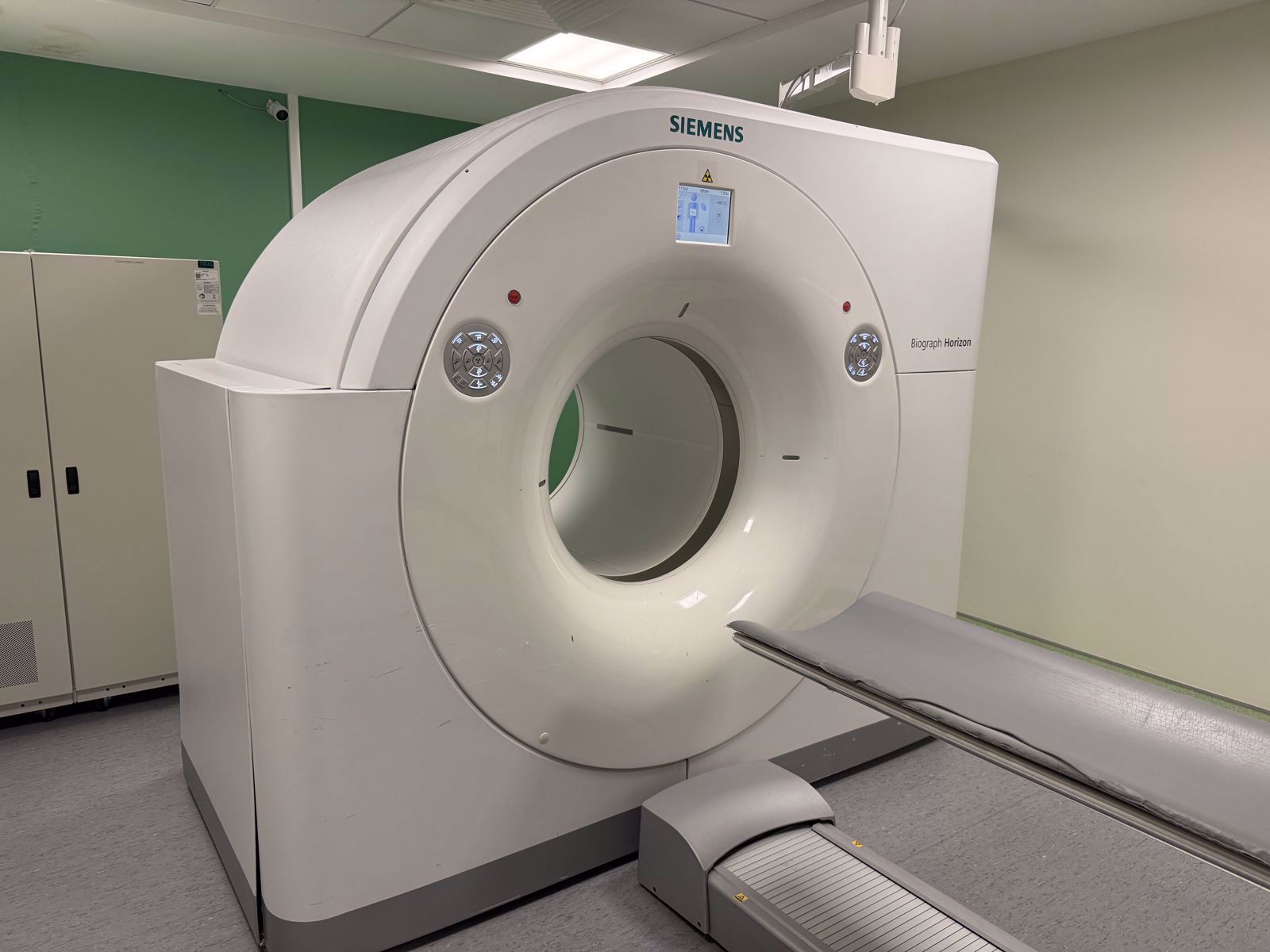
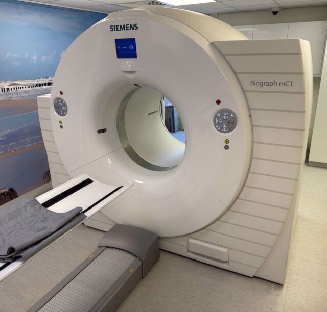
Key Components of a PET/CT Scanner
Understanding the components of a PET/CT scanner helps you evaluate systems more effectively—whether you are assessing a new installation or considering a pre-owned unit. The following are the major subsystems that define a PET/CT system's performance and reliability.
The PET Detector Ring
The PET detector ring is the heart of the PET subsystem. It consists of thousands of small scintillation crystals arranged in a ring around the bore. When a gamma photon strikes a crystal, the crystal produces a flash of light, which is then converted into an electrical signal by a photodetector.
The type of scintillation crystal used has a direct impact on image quality and scan speed. Older systems commonly use bismuth germanate (BGO) crystals, while modern systems frequently employ lutetium oxyorthosilicate (LSO) or lutetium-yttrium oxyorthosilicate (LYSO) crystals. LSO and LYSO crystals offer faster scintillation decay times, which translates into better timing resolution and improved image quality—especially for time-of-flight (TOF) PET imaging.
The CT X-Ray Tube and Detector Array
The CT subsystem of a PET/CT scanner contains the same core components found in a standalone CT scanner: an X-ray tube, a high-voltage generator, and a detector array. The X-ray tube is the most expensive and most frequently replaced component in any CT system. Therefore, understanding its heat capacity—measured in Mega Heat Units (MHU)—and its usage history is critical when evaluating any pre-owned PET/CT system.
The detector array determines how many slices the CT can acquire per rotation. PET/CT systems commonly feature CT components ranging from 16-slice to 128-slice configurations, depending on the generation and intended clinical use of the system.
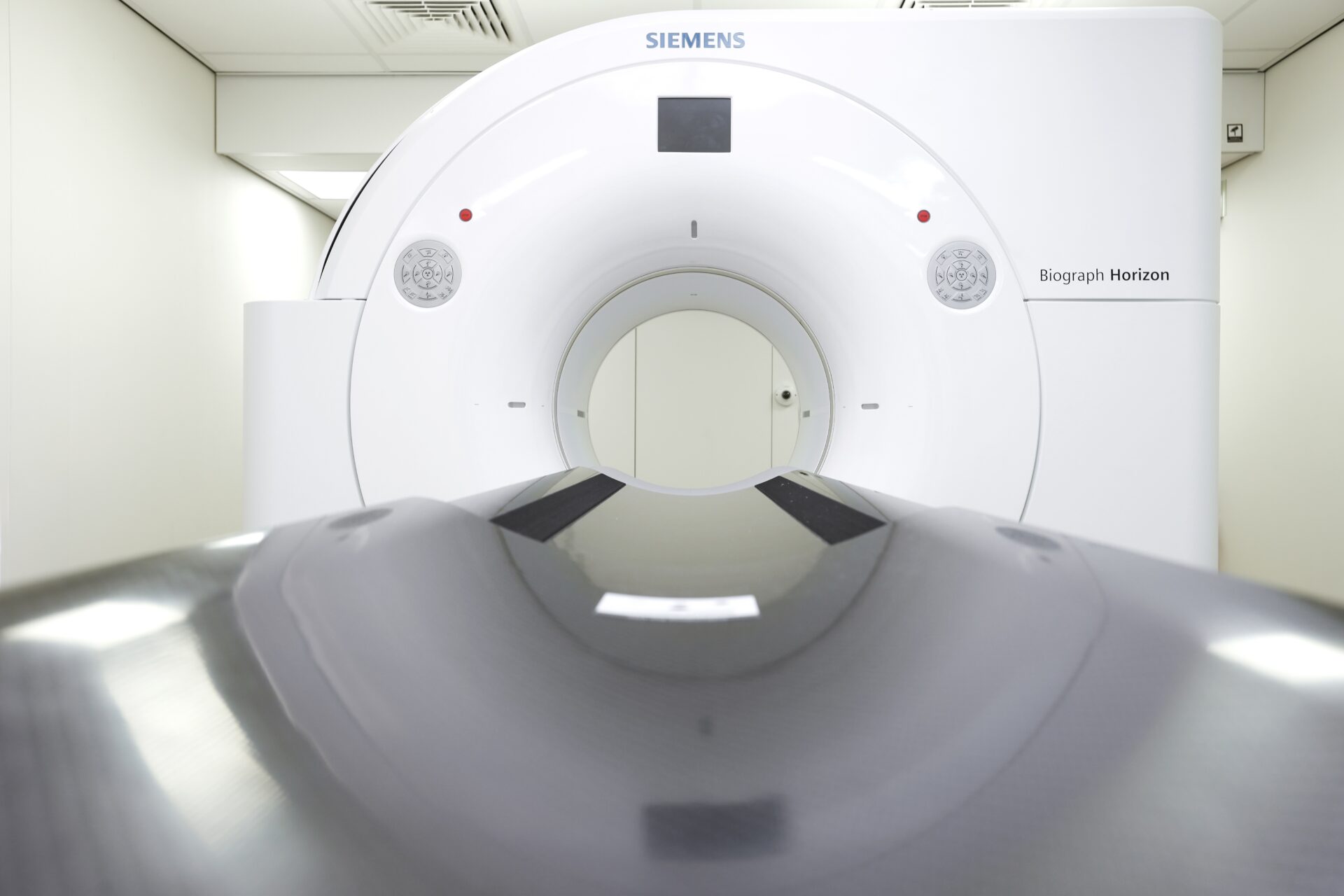
The Gantry and Patient Table
The gantry houses both the PET detector ring and the CT components. In most modern PET/CT systems, the CT section and PET section are positioned sequentially within a single housing, though they maintain separate detector systems. The patient table moves the patient through each section in a single continuous motion.
Table weight capacity, bore size, and axial travel range are all factors that affect clinical versatility. Systems designed for bariatric patients, for example, require wider bore diameters and higher table weight limits.
Acquisition and Reconstruction Workstation
The acquisition workstation controls the scan protocols, manages data acquisition from both the PET and CT subsystems, and performs image reconstruction. Modern systems use iterative reconstruction algorithms that deliver higher image quality at lower radiation doses compared to older filtered back-projection methods.
The workstation also handles the critical task of image fusion—aligning the PET and CT datasets so that the metabolic and anatomical information overlay with precision. Software capability at this level directly impacts diagnostic confidence.
Advantages of PET/CT Over Standalone Imaging
The decision to invest in a PET/CT scanner—rather than relying on separate PET and CT systems—carries significant clinical and operational advantages. The following are the most impactful benefits that drive adoption across healthcare facilities worldwide.
Superior Diagnostic Accuracy
The fusion of metabolic and anatomical data provides a level of diagnostic accuracy that neither modality can achieve alone. In oncology, PET/CT has been shown to improve tumor detection, staging accuracy, and the assessment of treatment response. The ability to localize a metabolically active lesion within precise anatomical structures reduces ambiguity and supports more targeted treatment planning.
Reduced Scan Time and Improved Patient Comfort
Because PET/CT acquires both datasets in a single session, patients do not need to be scheduled for two separate exams on two different machines. This reduces total scan time, minimizes patient discomfort, and eliminates the spatial registration errors that occur when PET and CT images are acquired on different days or different systems.
Streamlined Clinical Workflows
From an operational perspective, a single PET/CT system consolidates two imaging workflows into one. This simplifies scheduling, reduces the need for duplicate patient preparation, and allows your radiology department to manage throughput more efficiently. For facilities with limited floor space, the combined footprint of a PET/CT system also offers a practical advantage over maintaining two separate scanners.
CT-Based Attenuation Correction
As mentioned earlier, the CT component provides attenuation correction data for the PET images. This replaces the older method of using a rotating transmission source, which was slower and less accurate. CT-based attenuation correction is faster, more precise, and contributes to the overall reduction in scan time that PET/CT delivers.
Clinical Applications of PET/CT Scanners
PET/CT technology has established itself as indispensable across multiple clinical disciplines. While oncology remains the primary driver of PET/CT utilization, the technology's reach extends well beyond cancer diagnosis.
- Oncology: Tumor detection, staging, restaging, and monitoring treatment response across virtually all solid tumor types.
- Cardiology: Assessment of myocardial viability and perfusion, particularly in patients with coronary artery disease or heart failure.
- Neurology: Evaluation of neurodegenerative conditions such as Alzheimer's disease, as well as localization of epileptic foci for surgical planning.
- Infectious Disease: Identification and localization of occult infections and inflammatory processes, including fever of unknown origin.
- Radiation Therapy Planning: Precise delineation of tumor volumes for radiation treatment planning, improving the accuracy of targeted therapy.
The breadth of these applications underscores why PET/CT has become a cornerstone of modern diagnostic imaging departments.
Considerations When Acquiring a PET/CT System
Selecting the right PET/CT scanner involves balancing clinical requirements, budget constraints, and long-term operational costs. This is a complex decision, and it benefits from careful evaluation of several key factors.
Firstly, determine the primary clinical applications your facility needs to support. A system intended primarily for oncologic staging has different performance requirements than one used heavily for cardiac viability studies. Your clinical workflow should drive the specification, not the other way around.
Secondly, evaluate the CT component carefully. The number of slices, the X-ray tube's heat capacity, and the tube's usage history all affect both diagnostic capability and the system's total cost of ownership over time.
Lastly, consider whether a pre-owned PET/CT system could meet your clinical requirements at a significantly lower capital cost. High-quality pre-owned systems from leading Original Equipment Manufacturers (OEM) such as Siemens, GE, and Philips can deliver outstanding clinical performance when they have been properly sourced, tested, and maintained.
How Scandinavian Medical Solutions Supports Your PET/CT Needs
At Scandinavian Medical Solutions, we specialize in pre-owned diagnostic imaging equipment, spare parts, and flexible rental solutions across all major imaging modalities—including PET/CT. We work with systems from Siemens, GE, Philips, and Canon (Toshiba), and our team brings deep technical knowledge to every engagement.
We understand that acquiring or maintaining a PET/CT scanner is a decision with significant financial and clinical implications. That is why we approach every conversation as a partnership—listening to your clinical requirements, evaluating your operational context, and helping you find the right solution at the right price.
Whether you need a pre-owned PET/CT system, a critical spare part to minimize downtime, or a rental solution to bridge a gap in your imaging capacity, we are here for you. Do not hesitate to contact our team for guidance. You can reach us at +1 (714) 240-0864 or email us at sales@scandinavian-medical.com. We are your trusted partner in diagnostic imaging—ready to help you deliver the best possible care to your patients.